Introduction
Staff Details
Activities
Publications and Research
Technical Guidelines
Unit/Laboratories
Contact Us
Introduction
Centre for Arboviral and Zoonotic diseases
Mandate of the Division:
Centre for Arboviral And Zoonotic Diseases comprises of 9 National Reference Laboratory dealing with Zoonotic diseases of public health importance including outbreak prone and emerging infectious diseases. The role of the division is primarily to provide laboratory evidence by conducting special and reference level tests which are not available at most of the institutes or medical colleges in India. Currently the work is being carried out on the following Zoonotic diseases: Rabies, Kala-azar, Arboviral infections (Dengue, JE, Chikungunya, Zika virus & CCHF) Toxoplasmosis, Brucellosis, Leptospirosis, Rickettsiosis, Hydatidosis, Neurocysticercosis, Plague and Anthrax etc CAZD is also involved in Plague surveillance and preparedness assisted with Plague surveillance unit, NCDC Bengaluru Branch.Relevant Photograph of the Division


NCDC, DELHI, 2017

ZOONOTIC DISEASE PROGRAMMES, 2021


ORGANISED BY CAZD

KNOWN AS CENTRE FOR ARBOVIRAL AND ZOONOTIC DISEASES)


CONDUCTED BY CAZD IN COLLABORATION WITH RAMJAS COLLEGE ON
THE OCCASION OF “NATIONAL SCIENCE DAY”, 2023

MEDICAL COLLEGE & SMT. S.K. HOSPITAL, 2023
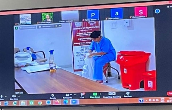
TRAINING ON PPE, SPILL MANAGEMENT & TRIPLE LAYER PACKING, 2019
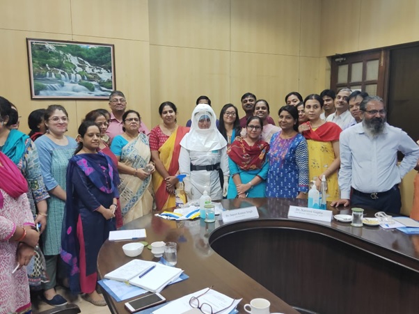
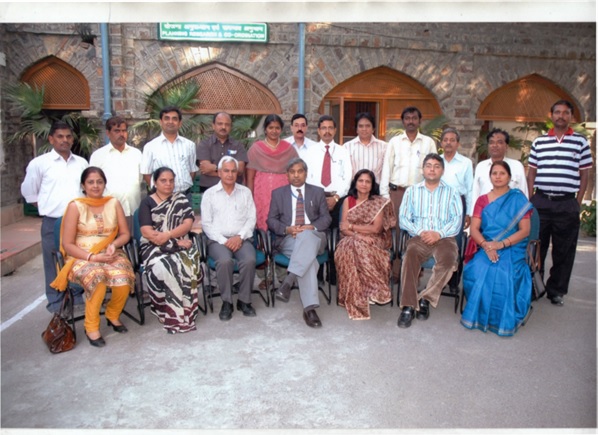
PHOTOGRAPH OF CAZD (FORMERLY ZOONOSIS DIVISION) OFFICIERS AND OFFICIALS, NCDC, DELHI, 2017
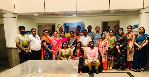
LABORATORY EXPERT GROUP MEETING FOR PROGRAMMES UNDER, DIVISION OF ZOONOTIC DISEASE PROGRAMMES, 2021
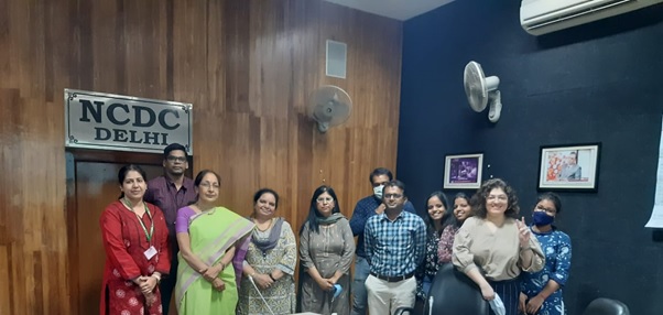
HANDS ON TRAINING ON DIAGNOSTIC RICKETTSIOLOGY, 2020
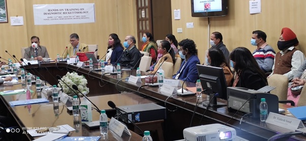
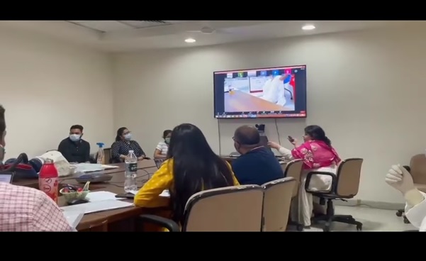
BIOSAFTETY AND BIOSECURITY WORKSHOP UNDER HISICON-2021, ORGANISED BY CAZD
OFFICIER AND OFFICIALS OF ZOONOSIS DIVISION FORMED IN 1963 (PRESENTLY KNOWN AS CENTRE FOR ARBOVIRAL AND ZOONOTIC DISEASES)
Staff Details
Staff Details
Division Officer’s

Dr. Monil Singhai
Joint Director
MBBS MD Microbiology
nicdzoonosis[at]yahoo[dot]com

Dr. Alka Singh
MBBS (LHMC)
Medical Officer
nicdzoonosis[at]yahoo[dot]com
Other Staff in the Division

Ms. Neeru Kakkar
Assistant Research Officer
M.Sc. (Bio. Chemistry)
N/A

Sh. R.K Pandey
M.Sc
Assistant Research Officer
N/A

Ms. Yosman
Assistant Research Officer
M.Sc. (Micro)
N/A

Ms. Niti Akoj
Assistant Research Officer
M.Sc. (Micro)
N/A

Dr. Rekha Jaiswal
Assistant Research Officer
M.Sc. (Phd)
N/A

Sh. Girrraj Singh
Research Assistant
M.Sc., M. Phil PGD.MPH PGD One Health
N/A

Ms. Sharda Singh
Research Assistant
M.Sc. (Zoo.) B. Ed
N/A

Sh. Chandan Singh
Research Assistant
B.Sc. (Biology)
N/A

Sh. K. S. Pandey
Research Assistant
M.Sc. (Zoo)
N/A

Sh. Vinay Singh
Technician
M.Sc. (Micro)
N/A

Ms. Jyoti
Technician
M.Sc. (Micro)
N/A

Ms. Preeti Khatri
Technician
M.Sc. (Biotech)
N/A

Ms. Neha Aggarwal
Technician
M.Sc. (Zoology)
N/A

Ms. Priyanka Yadav
Technician
M.Sc. (Micro)
N/A

Sh. Man Mohan Singh Mehra
Technician
10th Pass
N/A

Sh. Sumit Shukla
Technician
B.Sc (Biochemistry)
N/A

Ms. Saraswati Paeik
Lab Assistant
12th Pass
N/A

Sh. Balraj Singh
Lab Assistant
12th Pass
N/A

Sh. Parveen Kumar
Lab Assistant
10th Pass
N/A

Sh. Vinod Kumar
Insect Collector
12th Pass+1year computer
N/A

Sh. Deepak Kumar
Insect Collector
12th Pass with Science
N/A

Sh. Suresh Kumar
Insect Collector
8th Pass
N/A

Sh. Rakesh
Insect Collector
9th Pass
N/A

Ms. Babita Singhal
Insect Collector
B.Com
N/A

Sh. Madan Lal
Insect Collector
12th Pass
N/A

Sh. Devender Singh
Lab Attendant
10th Pas
N/A

Sh. Samar Nath
Lab Attendant
10th Pass
N/A
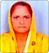
Ms. Rani
Lab Attendant
7th Pass
N/A

Sh. Naveen Kumar
Animal Attendant
10th Pas
N/A

Ms. Maya
Animal Attendant
N/A

Sh. Man Singh Meena
MTS
8th Pass
N/A

Ms. Monica
MTS
10th Pass
N/A
Activities
Publications and Research
Publications :
Research Papers- 1 Jayaprakasam M, Chatterjee N, Chanda MM, Shahabuddin SM , Singhai M,Tiwari S, Panda S. Human anthrax in India in recent times: A systematic review & risk mapping. One Health.Volume 16, June 2023, 100564. https://doi.org/10.1016/j.onehlt.2023.100564
- 2 Goyal R, Singhai M, Mahmood T, Saxena V. Association between the physical activity and metabolic syndrome in residents of a foot-hill area in India. Diabetes Metab Syndr. 2022 Mar 23;16(4):102471. doi: 10.1016/j.dsx.2022.102471
- 3 Agrawal SJ, Goyal R, Singhai M. Breakthrough Infections and Omicron Variant: Dealing with the Dilemma. Indian J Med Biochem 2021; 25 (3):0-0.
- 4 Singhai, M.; Sood, V.; Singh, G.; Siddiqui, C.; Nale, T.; Jha, P.; Yadav, P.; Jaiswal, R.; Bala, M.; Singh, S.K.; Tiwari, S. Rabies Outbreak in the Urban Area of Delhi: An Investigation Report and One Health Perspective for Outbreak Management. Infect. Dis. Rep. 2022, 14, 1033-1040.
- 5 Singhai M, Sood V, Yadav P, Kumar KK, Jaiswal R, Madabhushi S, Dhull P, Bala M, Singh SK, Tiwari S. Intravitam Diagnosis of Rabies in Patients with Acute Encephalitis: A Study of Two Cases. Infect Dis Rep. 2022 Nov 29;14(6):967-970. doi: 10.3390/idr14060095. PMID: 36547241; PMCID: PMC9778139.
- 6 Singhai, M., Shah, Y. D., Gupta, N., Bala, M., Kulsange, S., Kataria, J., & Singh, S. K. (2021). Chronicle down memory lane: India’s sixty years of plague experience. Indian Journal of Medical Microbiology.
- 7 Singhai M, Jaiswal R, Siddiqui C, Tiwari S, Gupta N, Bala M, Singh SK. Rabies can be a disease of puppyhood. J Family Med Prim Care. 2022 Jun;11(6):3339-3341. doi: 10.4103/jfmpc.jfmpc_1605_21. Epub 2022 Jun 30. PMID: 36119256; PMCID: PMC9480633.
- 8 Arora, R., Goel, R., Singhai, M., Gupta, N., & Saxena, S. (2021). Rabies Antigen Detection in Postmortem Cornea. Cornea, 40(5), e10-e11.
- 9 Singhai, M., Dhariwal, A. C., Gupta, N., Goswami, S., & Singh, S. K. (2020). An Observational Study of JE Cases Detected in National Centre for Disease Control from January 2017 to December 2019. Journal of Communicable Diseases (E-ISSN: 2581-351X & P-ISSN: 0019-5138), 52(4), 12-16.
- 10 Gupta, N., Singhai, M., Garg, S., Shah, D., Sood, V., & Singh, S. K. (2020). The missing pieces in the jigsaw and need for cohesive research amidst coronavirus infectious disease 2019 global response. medical journal armed forces india, 76(2), 132-135.
- 11 Jaiswal¹, R., Chhabra, M., Singh, P., Gupta, N., Singhai, M., Dhariwal, A. C., & Tripathi, P. (2018). Molecular Epidemiology and Sequence Analysis of Rabies Virus Isolates from North and North East India. Journal of Communicable Diseases, 50(4), 4
- 12 Singh, G., Chhabra, M., Singh, P., Gupta, N. K., Singhai, M., Dhariwal, A. C., & Ram, S. (2018). Molecular Study of Glycoprotein (G) Gene Region of Rabies Virus from Spotted Deer, Delhi, India. Journal of Communicable Diseases, 50(3), 3.
- 13 Singh, G., Jaiswal, R., Chhabra, M., Sood, Y., Gupta, N., Singhai, M., Tiwari, S., Dhariwal, A.C., Sharma, R. and Ram10, S. (2017). Evaluation of Direct Rapid Immunohistochemistry Test (DRIT) for Postmortem Diagnosis of Rabies. Journal of Communicable Diseases, 49(3), 3.
- 14 Rawat, V., Kumar, A., Kumar, M., Singhai, M., Rawat, C. M. S., & Jha, S. K. (2016). Viral Hepatitis A and E Outbreaks in Kumaon Region of Uttarakhand. National Academy Science Letters, 39(1), 17-19.
- 15 Singhai, M., Rawat, V., Singh, P., & Goyal, R. (2016). Fatal case report of concomitant hepatitis E and Salmonella paratyphi A infection in a sub-Himalayan patient. Annals of Tropical Medicine & Public Health, 9(1).
- 16 Malik, Y. P. S., Kumar, N., Rawat, V., Sharma, K., Kumar, A., Singhai, M., & Kumar, M. (2015). Detection and distribution pattern of prevalent genotypes of hepatitis-C virus among chronic hepatitis patients from Kumaon region of Uttarakhand, India. Indian journal of medical microbiology, 33, 161.
- 17 Mittal, V., Chhabra, M., & Venkatesh, S. (2015). Ebola virus-an Indian perspective. The Indian Journal of Pediatrics, 82(3), 207-209.
- 18 Sharma, P., Mittal, V., Chhabra, M., Singh, P., Bhattacharya, D., & Venkatesh, S. (2014). Dominance shift of DENV-1 towards reemergence and co-dominant circulation of DENV-2 & DENV-3 during post-monsoon period of 2012 in Delhi, India. Journal of Virology and Retrovirology, 1(1), 104.
- 19 Shrivastava, A., et al (2015). Outbreaks of unexplained neurologic illness—Muzaffarpur, India, 2013–2014. MMWR. Morbidity and mortality weekly report, 64(3), 49.
- 20 Sharma, P., Mittal, V., Chhabra, M., Kumari, R., Singh, P., & Venkatesh, S. (2016). Molecular epidemiology and evolutionary analysis of dengue virus type 2, circulating in Delhi, India. Virusdisease, 27(4), 400-404.
- 21 Singh, P., Chhabra, M., Sharma, P., Jaiswal, R., Singh, G., Mittal, V., Rai, A. and Venkatesh, S. (2016). Molecular epidemiology of Crimean-Congo haemorrhagic fever virus in India. Epidemiology & Infection, 144(16), 3422-3425.
- 22 Singh, P., Sharma, P., Kumar, S., Chhabra, M., Rizvi, M.A., Mittal, V., Bhattacharya, D., Venkatesh, S. and Rai, A. (2016). Continued persistence of ECSA genotype with replacement of K211E in E1 gene of Chikungunya virus in Delhi from 2010 to 2014. Asian Pacific Journal of Tropical Disease, 6(7), 564-566.
- 23 Thakur, R., Kumar, Y., Singh, V., Gupta, N., Vaish, V. B., & Gupta, S. (2016). Serogroup distribution, antibiogram patterns & prevalence of ESBL production in Escherichia coli. The Indian journal of medical research, 143(4), 521.
- 24 Kumar, Y., Gupta, N., Vaish, V. B., & Gupta, S. (2016). Distribution trends & antibiogram pattern of Salmonella enterica serovar Newport in India. The Indian journal of medical research, 144(1), 82.
Operational Research
- To study the epidemiological profile of Kala-azar patients in Delhi
- Serological studies in Toxoplasmosis in different Delhi Hospitals.
- Comparative analysis of various serological tests in diagnosis of Toxoplasmosis.
- Surveillance of Plague in different parts of the country.
- Molecular characterisation of strains of Leishmania.
- Standardization of appropriate diagnostic methods for sero-diagnosis and sero-epidemiology of leptospirosis.
- Surveillance of arboviral infections in man and animals.
- Study of prevalence of Rabies in peridomestic and wild rodents.
- Standardization of Rapid Fluorescent Focus Inhibition Test (RFFIT) for rabies antibody titer.
- Serological studies in clinically suspected cases of hydatid disease.
- Sero-epidemiological studies for rickettsial diseases (scrub typhus & Indian tick typhus) in patient with pyrexia of unknown origin.
Technical Guidelines
Technical Guidelines
Technical reports/Communicable Disease alerts:
- Glanders

- Kyasanur forest Disease: A Public Health Concern

- Meliodosis

- Scrub Typhus

- Zika Virus Disease

Manuals:
Unit/Laboratories
National Referral Laboratories/Units under Division:
| Sr. No | Lab/Unit name: |
|---|---|
| 1. | Plague/Anthrax Laboratory |
| 2 | Leishmania/Toxoplasma/ Cysticercosis/ Hydatid Laboratory |
| 3 | Arbovirus Laboratory (Dengue/Chikungunya/Japanese Encephalitis) |
| 4 | Leptospira/Brucella Laboratory |
| 5 | Rickettsia Laboratory /Borrellia Laboratory |
| 6 | Rabies Laboratory |
| 7 | Molecular Laboratory |
| 8 | Tissue Culture Laboratory |
| 9 | Quality control and Quality assurance |
| 10 | Central Plague Surveillance Unit |
A) ROUTINE REFERRAL DIAGNOSTIC SERVICES
| S.no | Disease | Name of the test | Turn Around Time * | Sample referral form(link) |
| 1. | Leishmaniasis (Kala-azar) | Bone marrow/liver/ spleen/ LN aspirate Staining for LD bodies | 1 day | Link for Leishmaniasis |
| Culture – Schneider medium | 10-14 days | |||
| Rk 39 RDT | 1 day | |||
| IFAt | 1-4 days | |||
| PCR for Leishmania donovani | 2-4 days | |||
| 2. | Toxoplasmosis | Toxoplasma Ig M ELISA | 1-4 days | Link for Toxoplasma |
| Toxoplasma Ig G ELISA | 1-4 days | |||
| Toxoplasma IG G Avidity ELISA | 1-4 days | |||
| PCR | 2-4 days | |||
| 3. | Cysticercosis | Ig G ELISA (T. solium) | 1-4 days | Link for Cysticercosis |
| 4. | Echinococcus | Ig G ELISA | 1-4 days | Link of Echinococcus |
| 5. | Dengue | Ig M ELISA for Dengue | 1-4 days | Link for Dengue |
| NS1 ELISA for Dengue | 1-4 days | |||
| NS1 RDT for Dengue | 1 day | |||
| Ig M RDT for Dengue | 1 day | |||
| Ig G RDT for Dengue | 1 day | |||
| PCR /QPCR for Dengue | 2-4 days | |||
| PCR /QPCR for Dengue Type specific | 2-4 days | |||
| 6. | Chikungunya | Ig M ELISA for Chikungunya | 1-4 days | Link of Chikungunya |
| Ig M RDT for Chikungunya | 1 day | |||
| Ig G RDT for Chikungunya | 1 day | |||
| PCR /QPCR for Chikungunya | 2 -4 days | |||
| 7. | Japanese Encephalitis | Ig M ELISA for JE | 1-4 days | Link for JE |
| QPCR | 2 -4 days | |||
| 8. | Rabies virus | IgG ELISA Rabies antibody detection in Human serum and plasma | 1-4 days | Link for antibody titre (Human) |
| Ig G ELISA Rabies antibody detection in animal (dogs/cats) serum and plasma | 1-4 days | Link for antibody titre (Animal) | ||
| RDT Antigen Detection in animal brain tissue (Canine/bovine/raccoon dog) | 1 day | Link for Proforma for a case of Hydrophobia (Human) | ||
| FAT for postmortem human/animal samples | 1 day | Link for Advisory and Proforma for Postmortem Sample sent for Medicolegal Purpose for Confirmation of Rabies | ||
| RT-PCR for human/animal samples | 2-4 days | Link for Epidemiology of cases of rabies in dogs & other animals | ||
| 9. | Brucellosis | Ig M ELISA for Brucella | 1-4 days | Link for Brucella |
| Ig G ELISA for Brucella | 1-4 days | |||
| B. abortus tube agglutination test | 1-4 days | |||
| B. melitensis tube agglutination test | 1-4 days | |||
| 10. | Leptospirosis | Ig M for Leptospirosis | 1-4 days | Link of Leptospira |
| Ig G for Leptospirosis | 1-4 days | |||
| 11. | Rickettsial diseases | Weil felix test-Ox2 | 1-4 days | Link for Rickettsial Diseases (Scrub Typhus) |
| Weil felix test-Ox19 | 1-4 days | |||
| Weil felix test-OxK | 1-4 days | |||
| Ig M ELISA for Scrub typhus | 1-4 days | |||
| Ig G ELISA for Scrub Typhus | 1-4 days | |||
| 12. | Lyme | ELISA | 1-4 days | Link for lyme |
| QPCR | 2-4 days |
Notes:
*For Samples received on working days (Monday-Wednesday) 10 am to 5 pm; turnaround time is tentative subject to availability of optimum batch size for complete utilization of kits Kindly contact concerned official/officer for information required regarding of case Investigation form, sample type, time, collection and transport. kit availability etc. before sending the samples Dr. Monil Singhai, JD /Dr. Alka Singh, MO, CAZD Contact No. 011-23913811, 011-20832482 Email id: nicdzoonosis@yahoo.com| S.no | Name of the Disease | Name of the test | Turn Around Time |
| 1. | CCHF | Ig M ELISA for CCHF | 1 day |
| Ig G ELISA for CCHF | 1 day | ||
| QPCR | 2-4 days | ||
| 2. | Dengue | Ig G ELISA for Dengue | 1 day |
| 3. | Chikungunya | Ig G ELISA for Chikungunya | 1 day |
| 4. | Zika Virus Disease Link for Zika proforma | Ig M ELISA for Zika | 1 day |
| Ig G ELISA for Zika | 1 day | ||
| RT-PCR /QPCR | 2-4 days | ||
| 5. | Brucellosis | Staining /Culture and Sensitivity | 7-10 days |
| 6. | Leptospirosis | RDT for Leptospirosis | 1 day |
| Microscopy: Dark field microscopy /Silver staining | 1-4 days | ||
| Culture on EMJH | 7-10 days | ||
| PCR for Leptospirosis | 2-4 days | ||
| 7. | Rickettsial Diseases | IFAT for Scrub Typhus | 1-4 days |
| IgM for Spotted Fever | 1-4 days | ||
| IgG for Spotted Fever | 1-4 days | ||
| IgM for Typhus Group | 1-4 days | ||
| IgG for Typhus Group | 1-4 days | ||
| PCR for Scrub Typhus | 2-4 days | ||
| 8. | Hantaan Virus | Ig M ELISA for Hantaan virus | 1 day |
| Ig G ELISA for Hantaan virus | 1 day | ||
| QPCR | 2-4 days | ||
| 9. | Chandipura Virus | QPCR | 2-4 days |
| 10. | KFD | QPCR | 2-4 days |
| 11. | Ebolavirus Disease (Zaire) | Ebolavirus Antigen ELISA | 1 day |
| QPCR | 2-4 days | ||
| 12. | Nipah Virus Disease | Ig M ELISA for Nipah | 1 day |
| Ig G ELISA for Nipah | 1 day | ||
| Nipah Antigen ELISA | 1 day | ||
| QPCR | 2-4 days | ||
| 13. | Monkey pox virus | QPCR | 2-4 days |
| 14. | Ortho pox virus | QPCR | 2-4 days |
| 15. | West nile virus | QPCR | 2-4 days |
| 16. | Yellow fever | QPCR | 2-4 days |
| 17. | Plague | Microscopy: Gram staining, Wayson staining | 1 day |
| Culture | 4-7 days | ||
| PCR | 2-4 days | ||
| 18. | Anthrax | Microscopy: Gram staining, Capsule staining | 1 day |
| Culture | 4-7 days | ||
| PCR | 2-4 days |
Contact Us
Contact Us
Full Mailing Address:
Centre for Arboviral and Zoonotic Diseases (CAZD), National Centre for Disease Control, Ministry of Health and Family Welfare
www.ncdc.gov.in
011-20832481
N/A


